Liver rupture leads to hemophilia B diagnosis in newborn: Case report
Infant made full recovery thanks to early detection, timely surgery
Written by |
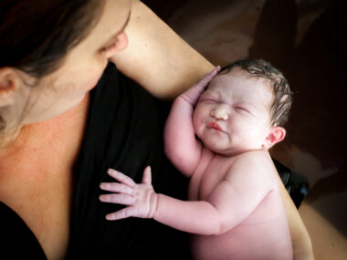
A newborn in China was diagnosed with hemophilia B after a ruptured liver caused significant abdominal bleeding, according to a case report. Due to early detection, timely surgical repair, and immediate factor-replacement therapy, the infant made a full recovery.
This case shows that “neonatal hepatic rupture may be the initial manifestation of an underlying bleeding disorder such as hemophilia B,” according to researchers. Newborns with spontaneous hepatic rupture should undergo testing to rule out coagulation disorders, they added.
The case was described in “Hepatic rupture in a neonate with hemophilia B: a case report,” published in the Journal of Pediatric Surgery Case Reports.

Hemophilia B occurs when the body lacks functional factor IX
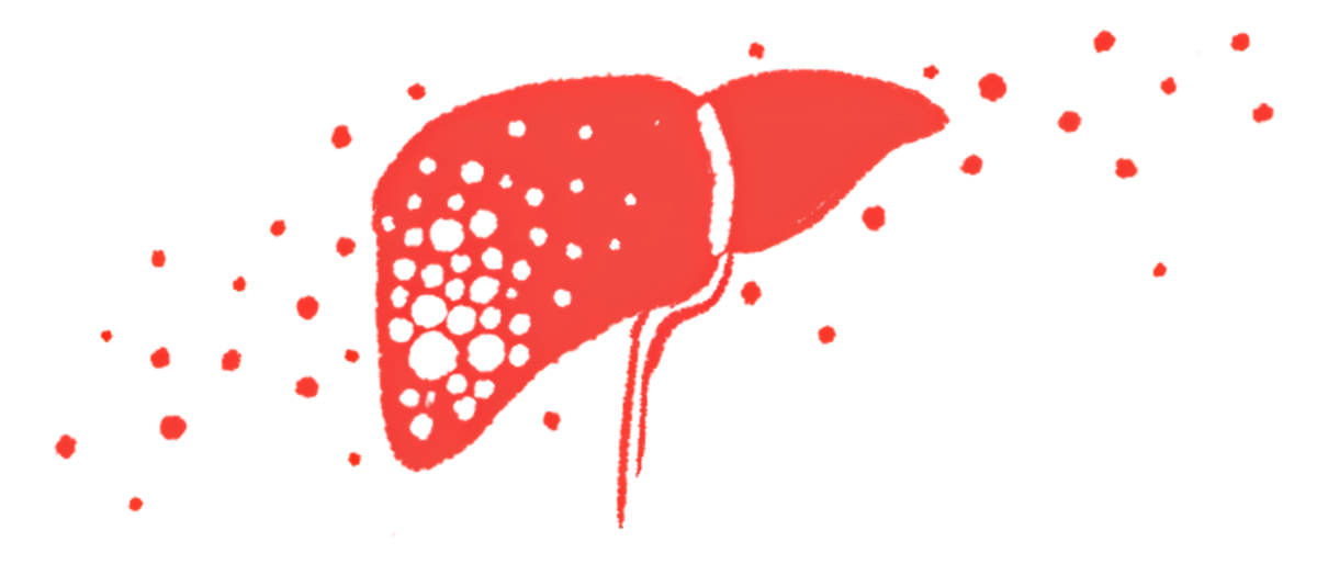
Hemophilia is a bleeding disorder caused by missing or malfunctioning clotting factors — the proteins that help blood clot properly. Hemophilia B occurs when the body lacks functional factor IX (FIX), usually due to mutations in the F9 gene.
In this report, researchers in China described the case of a newborn diagnosed with hemophilia B after developing intra-abdominal bleeding due to a liver rupture.
The boy was born via cesarean section at 39 weeks of pregnancy, with a birth weight of 3.6 kg (7.9 pounds). At 59 hours of life, he developed progressive abdominal swelling, without vomiting or fever, and his condition rapidly deteriorated as he exhibited symptoms of pallor, cold extremities, groaning, and lack of energy.
At admission, his temperature was 35 degrees C, and he had low blood pressure and low levels of hemoglobin, the protein that carries oxygen in red blood cells. He also had severe metabolic acidosis, a life-threatening condition where blood pH drops due to too much acid, with a pH of 6.57 and elevated lactate levels.
An ultrasound combined with paracentesis, a medical procedure to drain excess fluid from the abdomen, confirmed bleeding into the peritoneal cavity — the space within the abdomen that contains different organs, including most of the liver.
According to the researchers, “unexplained abdominal distension or pallor should alert clinicians to possible intra-abdominal hemorrhage. Bedside ultrasound and diagnostic paracentesis are invaluable tools for early detection.”

Boy made uneventful recovery from liver rupture surgery
An emergency surgery was performed on the newborn 63 hours after birth to assess the abdominal cavity. The researchers found a blood clot extending from the liver into the abdominal cavity and active bleeding from a 3 cm liver fissure, which was surgically repaired.
After the surgery, the boy received a transfusion of red blood cells, plasma (the liquid component of blood), platelets (cell fragments that help the blood clot), and concentrated coagulation factors. He was discharged in stable condition after two weeks, and made an uneventful recovery.
Coagulation function assays revealed a lower activity of FIX (2 %), leading to a diagnosis of hemophilia B. At 6 months of follow-up, he had no recurrent bleeding episodes and remained clinically stable on regular factor replacement therapy.
“This case emphasizes the link between coagulation disorders and solid organ rupture in neonates. Hepatic rupture without significant trauma should raise suspicion for an underlying bleeding [disorder],” the researchers wrote.