
MRI Superior to Radiography in Capturing Joint Changes That Signal Future Bleeds, Study Says


Joint changes detected by magnetic resonance imaging (MRI), and often are missed on radiographs, are a strong predictor of damaging bleeds over the next five years in joints of people with hemophilia, a study suggests.
A 10-fold increase in the five-year risk of joint bleeds was found by researchers in joints (ankles and knees) that appeared normal on radiographs but showed areas of thickened tissue on MRIs. These joints bled sooner and more often, and were prone to more severe injuries.
Patients should be monitored regularly for potentially damaging bleeds, they advised.
The study “MRI predicts 5-year joint bleeding and development of arthropathy on radiographs in hemophilia” was published in the journal Blood Advances.
Clotting factor replacement therapy, either given as a preventive treatment (prophylaxis) or on demand, is a mainstay in managing hemophilia. While effective, it cannot always prevent bleeds that often affect the joints, particularly the knees, ankles, and elbows.
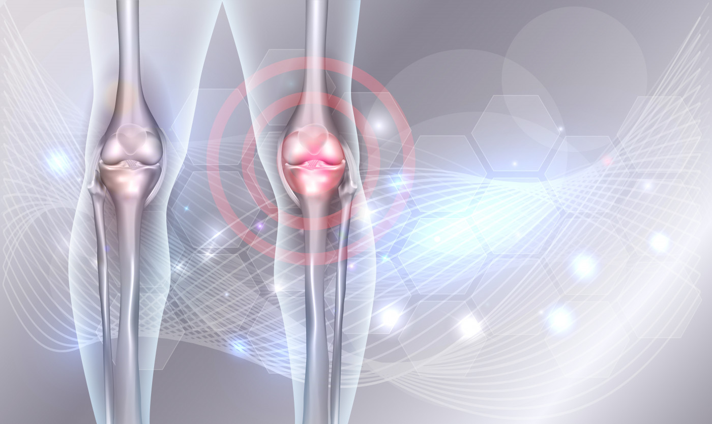
A common consequence of joint bleeding is synovial hypertrophy, or the enlargement (thickening) of the synovium — the connective tissue that lines the inside of the joint capsule. Such hypertrophy can result in cartilage destruction and degenerative changes in bones, ligaments, and joint capsules.
It also increases the risk of joint bleeds, which are progressively damaging. Arthropathy, or joint disease, is a common and disabling problem among people with bleeding disorders, and one that needs to be monitored.
Standard radiographs show advanced bone alterations, but usually at stages where these changes are irreversible. MRI is considered a gold standard for detecting early, even pre-symptomatic, changes to joints, even if the clinical relevance of early MRI findings is still not fully understood.
Researchers at University Medical Center Utrecht, in the Netherlands, assessed if early MRI results could predict the risk of joint bleeds and damage over the next five years in people with hemophilia seen at their clinic.
Both knees and ankles of 26 patients, ages 12 to 29 — all showing minimal or no joint disease on radiographs — were scanned by 3T MRI.
Nearly all patients with severe hemophilia (15 of 16; 94%) and one of eight with moderate hemophilia (13%) were receiving prophylaxis. None had inhibitors, meaning antibodies that render the treatment ineffective.
At the study’s beginning, the annualized bleeding rates in patients’ knees and ankles were low, a median of 0.2.
Radiography detected alterations in 7% of joints imaged, while MRI showed abnormalities in 39% of them, confirming that MRI is more sensitive in detecting joint changes.
The most common MRI findings were effusion (an abnormal accumulation of fluid in a joint; 23%), synovial hypertrophy (16%), and hemosiderin deposits (a buildup of iron-storage particles caused by the breakdown of hemoglobin during bleeds; 16%).
Over the next five years (follow-up period), the majority of patients (88%) reported bleeds in 36% of their knees or ankles. This figure did not differ between those with severe hemophilia on prophylaxis, and patients with moderate disease treated on demand.
MRI-detected synovial hypertrophy was a “strong and independent” predictor of future bleeding, the study noted.
Bleeding occurred more often in joints where synovial hypertrophy had been detected by MRI five years before, compared to joints where it was not evident — a median of two bleeds during these five years and none in healthier joints.
The five-year bleeding risk was 80% for joints with synovial hypertrophy and 27% for those without it, reflecting bleeding odds 10 times higher in joints with synovial problems.
All MRI-detected changes, except effusion, were also strong predictors of joint disease (arthropathy) developing five years later.
“In conclusion, MRI evaluation of joints provides relevant information in patients with hemophilia with absent or limited arthropathy. Synovial MRI changes were a strong predictor for 5-year bleeding and progression of arthropathy,” the researchers wrote.
“These findings,” they added, “emphasize the importance of monitoring joint status and may be used to guide treatment.”