
Implanting 3D Bioscaffolds in Rats Works to Trigger FVIII Production


Implanting bioscaffolds — 3D biological scaffolds made up of collagen and other molecules — carrying transformed liver cells into rats successfully triggered the production of factor VIII (FVIII), the clotting protein missing in people with hemophilia A.
According to researchers, these bioscaffolds may be a therapeutic alternative to FVIII replacement therapy, a current standard of care treatment for hemophilia A.
The new treatment strategy was demonstrated in a study titled “Implanted subcutaneous versus intraperitoneal bioscaffold seeded with hepatocyte-like cells: functional evaluation,” published in the journal Stem Cell Research & Therapy.
Replacement therapy is a standard treatment for hemophilia A in which the missing FVIII is supplied to patients either on an on-demand basis, to treat bleeds, or on a prophylactic basis, to prevent them.
However, replacement therapy comes with certain disadvantages, including life-long dependence and high costs. Patients using this therapy also may develop antibodies that target the delivered FVIII, reducing its effectiveness.
Because FVIII is normally produced by liver cells (hepatocytes), one potential long-term therapeutic strategy for hemophilia A patients is implanting stem cells that can grow into healthy FVIII-producing liver cells.
One method of delivering these stem cells is to implant them onto the liver’s extracellular matrix (ECM), a collection of proteins such as collagen and other molecules that form a 3D scaffold that surrounds and supports cells. Existing cells can be removed from the liver’s ECM — through a process known as decellularization — and replaced with stem cells that become fully functioning liver cells.
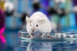
Now, researchers at Cairo University, in Egypt, and their colleagues explored the feasibility of this method by transplanting a decellularized liver ECM, or bioscaffold, seeded with human stem cells, into rats. Their goal was to determine if human stem cells would transform, or differentiate, into liver cells capable of making FVIII.
The ECM scaffolds were isolated from pigs, decellularized, and then seeded with a type of adult stem cells known as mesenchymal stem cells, which were extracted from human bone marrow. These stem cells were treated for 14 days with two proteins that induced their differentiation into liver-like cells.
Microscope analyses allowed researchers to determine whether or not the stem cells had transformed into cells with liver-like characteristics by examining their appearance: differentiated cells tended to form clusters with distinct shapes and round nuclei.
The cells were then stained to identify structural and molecular features characteristic of differentiated liver cells. By day seven of the differentiation process, the cells began expressing, or producing, alpha-fetoprotein (AFP), a marker of immature liver cells. On day 14, the expression of AFP had decreased, but that of cytokeratin 19 (CK19), a marker of mature liver cells, had increased.
Similarly, the area covered by AFP-producing cells expanded by day seven and then significantly decreased by day 14. Moreover, the area of cells expressing CK19 continued to increase throughout the 14-day differentiation process.
Cell differentiation was confirmed by the significantly increased production of the protein albumin, which is produced by liver cells, compared with undifferentiated control cells (mean of 1.06 vs. 0.2 mcg/mL). AFP’s mean level also was significantly higher in differentiated cells than in control cells (mean of 20.8 vs. 12.93 mcg/mL).
To evaluate the seeded bioscaffolds in vivo, or in a living organism, the researchers implanted them into 14 rats: seven under the skin, or subcutaneously, and seven into the abdomen, called intraperitoneally. Seven animals that did not receive any form of treatment were used as controls. After 10 days, blood samples, as well as skin and peritoneal tissues, were collected from all animals to be analyzed.
Blood tests confirmed the production of human FVIII in animals that received the seeded bioscaffolds, with rats treated subcutaneously showing significantly higher levels than those treated intraperitoneally.
“Increased mean values of [blood] FVIII in groups subjected to subcutaneous implantation make subcutaneous more preferable than intraperitoneal one,” the researchers wrote.
An examination of skin sections from rats treated subcutaneously revealed the presence of transformed liver-like cells at the base of hair follicles that also were positive for AFP and CK19. Likewise, liver-like cells positive for these markers also were seen in peritoneal tissue sections from rats treated intraperitoneally.
The team also found the area of immature AFP-positive cells was greater in peritoneal tissue samples compared with skin sections. In contrast, the area of mature liver-like cells expressing CK19 was significantly higher in skin tissue than in peritoneal sections.
“Implantation of decellularized bioscaffold seeded with [differentiated] stem cells in rats was successful to establish production of FVIII,” the researchers wrote.
“Decellularization strategy for synthesis of bioscaffold seeded with hepatocyte-like cells has the potential to present an alternative for recombinant factor VIII replacement to treat hemophilia patient,” they concluded.