Bone Defects Detected at Birth, After Injury, in Mouse Models of Hemophilia A and B, Study Shows

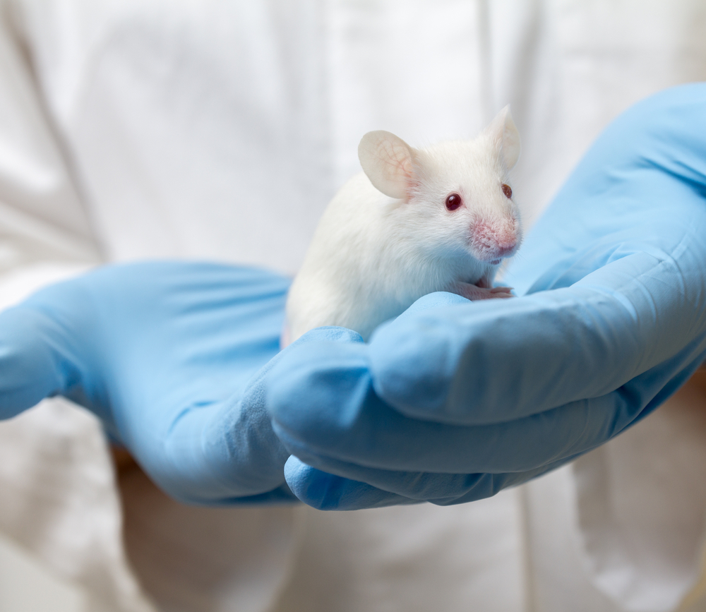
Bone defects were seen since birth — and following injury — in mouse models of hemophilia A and B, but not in Von Willebrand (VWD) disease mice, a study shows.
Researchers said further study into the potential mechanisms of primary bone deficits in hemophilia may help in developing screening and treatment guidelines.
The study, “Hemophilia A and B mice, but not VWF−/−mice, display bone defects in congenital development and remodeling after injury,” was published in the journal Scientific Reports.
Severe hemophilia is associated with chronic degenerative joint and bone disease, but patients also show signs of osteopenia, or bone loss, and osteoporosis — reduced bone density.
Von Willebrand disease, called VWD, is a common, inherited hemorrhagic disorder. It results from a mutation in the gene that codifies a protein called von Willebrand factor (VWF). This protein circulates in the blood and helps platelets adhere to sites where the blood vessel has been injured, also carrying and stabilizing the procoagulant protein factor VIII (FVIII).
Mutations in the VWF gene impair the proper interaction between platelets and the vessel wall, interfering with hemostasis, which is the process that causes bleeding to stop.
To learn more about the effects of these diseases on bones, the researchers evaluated bone health in mouse models of hemophilia A, hemophilia B, and VWD. The mouse model for hemophilia A lacked the gene coding for FVIII, while the hemophilia B mice lacked the FIX gene. The VWF mice lacked the VWF gene. These mice are called knockout mice.
To assess the bone’s capacity to regenerate, the investigators injured the animals’ knees. The mice’s bone mineral density (BMD) was evaluated both before and after injury.
Compared with control mice — called wild-type — the hemophilia A and hemophilia B mice showed defects in bone mineral density and bone mineral content before any injury was inflicted. Bone mineral density was 3.44% lower in the hemophila A mice, and 4.54% lower in the hemophilia B mice at 16 weeks of age, compared with the controls. No differences were seen in the mouse model of VWD disease.
Analysis of the shinbone (proximal tibia) at the age of 22 to 24 weeks showed a volume loss of bone mineral density of 27.8% in the hemophilia A mice and of 15.5% in the hemophilia B mice compared with controls.
Researchers then inflicted an injury to the knee joint and assessed the outcomes after two weeks. The analysis showed that, in the controls, the joint went back to normal. In comparison, the joints of the hemophilia A and B mice remained inflamed, as shown by high scores — 5.57 and 5.18 — on the Valentino scale, a zero-to-10-point scale that measures damage of the joints in mice. Higher scores mean increased damage. The score of the injured mouse model of VWD was 0.95.
Further analysis revealed significant changes to the injured bone in both hemophilia mice, including increased porosity and surface roughness compared with the non-injured limb or control mice.
These changes were accompanied by increased levels of a pro-inflammatory cytokine, or signalling molecule, called interleukin-6 (IL-6). There also was an increase in the number of osteoclasts — cells that absorb bone tissue during growth and healing — and significant loss of the trabecular bone, also called spongy bone.
Researchers continued to observe disorganized bone formation and resorption, which caused significant deterioration of the joint in the hemophilia mice.
“Mice with complete FIX or FVIII deficiency (but not VWF deficiency) display similarly defective congenital [since birth] bone homeostasis [balance] and bone remodeling following hemarthrosis [bleeding into the joints],” the researchers said.
“Further study of potential mechanisms of primary bone deficits in hemophilia is needed to guide appropriate screening and treatment guidelines,” the study concluded.


