
Brief electrical pulse may help in hemophilia gene therapies: Study
In lab, liver cells take up viral carrier more easily with stimulation
Written by |

Gentle electrical stimulation may help liver cells uptake gene therapies, such as those used to treat hemophilia, according to new research by a team of U.S. scientists.
In lab studies, the team found that liver cells exposed to a brief electrical field more easily took up a viral carrier, like the ones used in gene therapies, as compared with unstimulated cells.
The researchers believe this approach could ultimately lower the necessary dose of these gene therapies, helping to lower treatment costs and make them safer.
“These results have important implications for gene therapy research, as they suggest that [electric pulsing] and optimized conditions can lead to improved gene delivery with significantly reduced AAV [carrier virus] dosages,” the researchers wrote.
The study, “Electric pulse exposure reduces AAV8 dosage required to transduce HepG2 cells,” was published in PLOS One.

Scientists’ goal is to reduce AAV dose needed for gene therapies
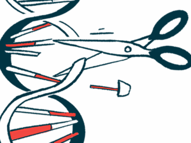
Gene therapy has emerged as a promising approach for treating diseases with a known genetic cause. It involves adding, editing, or removing genes to compensate for the genetic defect — thus offering a possibly curative intervention.
To date, three gene therapies have been approved for treating hemophilia, in which genetic defects result in the lack of specific factors needed for proper blood clotting.
Roctavian works to provide a functional version of F8, the gene that’s faulty in hemophilia A, while Hemgenix and Beqvez provide a working version of F9, the gene implicated in hemophilia B.
Each is packaged into a viral carrier, or vector, called an adeno-associated virus or AAV. This helps it be taken up by a patient’s liver cells, where blood clotting proteins are mainly produced, when given as an infusion into the bloodstream.
Viruses are very good at incorporating themselves into human cells, so scientists use versions of them that won’t cause disease as a way of delivering genetic material.
It’s important, the researchers note, to get the right dose of a gene therapy into the patient. If too little is infused, target cells might not take up enough of it and the therapeutic effects may be limited. Conversely, delivering too much of the viral vector could cause a person’s immune system to react, leading to safety issues, in addition to driving up costs.
“Lowering the required AAV dose could significantly improve safety and reduce the cost of AAV-vector-mediated, liver-directed gene therapies,” the researchers wrote.
Over the last decade, scientists have been working to develop ways to achieve this.
Lowering the required AAV dose could significantly improve safety and reduce the cost of AAV-vector-mediated, liver-directed gene therapies.
Previous research had shown that exposure to an electrical field makes it easier for molecules to pass through a cell’s external cell membrane and reach its interior. For a gene therapy, that could possibly mean that even when a lower dose is infused, enough of it can be taken up by liver cells to have the same therapeutic effect.
Researchers tested approach using green fluorescent protein
Here, a team led by researchers at the University of Wisconsin-Madison looked at “a way of getting the treatment DNA directly into the liver without passing it through the entire body and triggering the immune system,” Susan Hagness, PhD, a professor at UW-Madison and a study author, said in a university press release.
“What we started talking about was … whether we could use electric pulses in order to make this delivery process more efficient and dramatically reduce the dose needed,” Hagness said.
The researchers conducted laboratory experiments to determine whether human liver cells grown in the lab would more readily take up gene therapy virus particles if they were first exposed to an electrical field.
Cells were either left unstimulated or given a brief electrical pulse before being incubated with AAV particles containing a green fluorescent protein as their payload. This protein could be visualized to see how readily the viral particles were taken up by liver cells.
The results showed that the electrically stimulated cells took up substantially more fluorescent protein compared with unstimulated cells.
Overall, the electrical pulses reduced the AAV dosage needed for the same amount of fluorescent protein to be taken up by liver cells — called transduction efficiency — by more than 50 times, according to the researchers. Moreover, they did not cause lasting damage to the cells.
The scientists also conducted a handful of experiments to understand exactly how the electrical stimulation works, especially since virus particles “already have their own way of getting inside cells,” according to John Booske, PhD, now a professor emeritus at UW-Madison and the study’s senior author. Still, they haven’t come up with a firm conclusion on the mechanism yet.
The team now is pursuing additional funding to continue studying this approach for improving gene therapy delivery. They hope it will eventually reach clinical trials, “reducing risks to the patient and improving efficacy” of virus-mediated gene therapy, the team concluded.


Leave a comment
Fill in the required fields to post. Your email address will not be published.